


 202-203 / 568

Characterization of Microcirculatory Response to
Gravity-Induced Changes using Noninvasive Thermal Imaging
Noam Moyal, Noa Darchi
Advisors: Dr. Zehava Blechman
Consultant: Dr. Oshrit Hoffer , Dr. Neta Rabin, Dr. Benjamin Gavish.
Medical Engineering
This research project focuses on analyzing hemodynamics microcirculation changes during
gravitation conditions using noninvasive techniques. This multidisciplinary research project
focuses on hemodynamic changes in peripheral microcirculation during gravitational
conditions in non-invasive techniques. The study was based on a clinical trial and included the
establishment of a trial system and physical measurements, as well as an data analysis
including digital image processing and machine learning.
Population:
The study was included 30 healthy volunteers, without any known
vascular pathologies, in 3 age groups: 15-49y ,50-64y, 65y and over. The clinical trial
was approved by Afeka Ethics committee (ID 05-04-2017-1-AFK)
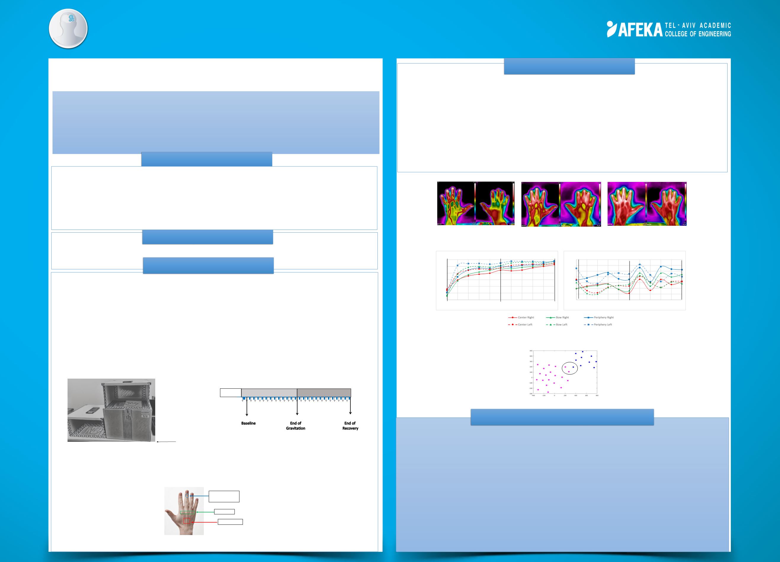
Experimental system
(Figure 1)
:
A user-friendly system was established in order to
induce palm gravity conditions. The system is also portable, and thus allows to
perform the experiment at subjects' homes, making it easier for the older subjects.
Throughout the experiment, thermal images were captured using 2 Flir one® pro
Compact Thermal Cameras and 2 smartphones. Figure 2 illustrates the experimental
protocol.
Thermal image processing:
3 Sub-regions in each hand were determined according
to the anatomical blood flow. Mean ST were calculated at 3 areas using Flir Tools
software. (Figure 3).
To characterize the microcirculatory response of gravity-induced changes using
thermal imaging.
Background
The increasing of palm skin temperature during gravity conditions reflects blood perfusion
compensation due to high local oxygen consumption, while local blood pressure decreases.
The bilateral effect indicates the Central Nervous System (CNS) involvement.
Thermal imaging allows non-invasive quantitative characterization of the palm’s blood
distribution under gravitational conditions.
Thermal monitoring of blood supply may promote new aspects of hemodynamics research.
Implementation of the study's insights could lead to improved the assessment of blood
supply in clinics, as well as at home, based on patient-specific diagnosis.
Gravitation caused ST increase in both palms, that began with one hand elevating and
remained high during the recovery period (Figure 4).
Different patterns of ST changes were observed in different subjects : While an increase in
temperature and stabilization was observed in some subjects (Figure 5, left), others showed
pulsations which describing an attempt of stabilization (Figure 5, right).
K means algorithm divided the subjects into two different groups (Figure 6) . The first one
characterized by lower initial temperatures and significant temperature differences between
the palm’s center and fingertips , while the second group characterized by higher initial
temperatures, and there are almost no temperature differences in the palm’s areas.
Methods
Conclusions & Applications
Objectives
Results
Peripheral microcirculatory
has been already found to reflect both local and systemic changes
during different conditions of perfusion and even predict the development of ischemic stress
conditions.
Infrared Thermal Imaging
is a noninvasive, non-contact and safe technique method to
measure skin temperature (ST), which has been proven in our laboratory as sensitive for
diagnosing changes in peripheral blood flow.
Figure 1.
The experiment system
Heart level
Figure 2.
Experimental protocol
Thermal image
every 10 seconds
Gravitation (10 minutes)
Recovery (10 minutes)
Baseline
B
A
C
Figure 4
. The thermal images of both palms at baseline stage (A), following 10
minutes gravitation (B) and 10 minutes recovery (C)
25
27
29
31
33
35
37
0
2
4
6
8
10
12
14
16
18
20
Temperature [
℃
]
Time [Min]
No
8
- Age
26
33
33.5
34
34.5
35
35.5
36
36.5
0
2
4
6
8
10
12
14
16
18
20
Temperature [
℃
]
Time [Min]
No
29
- Age
77
Figure 5
. Change in ST for different anatomical regions of both palms at young
participant (26 years old) and at old participant (77 years old)
Distal phalanx of
the middle finger
Palm Bow
Palm Center
Figure 3.
Three different areas
Figure 5
. Two groups by k means algorithm